PRP Therapy
- Home
- PRP Therapy
Platelet-Rich Plasma (PRP) Therapy: A Powerful autologous regeneration solution
Platelet-rich plasma (PRP) is a highly effective treatment that uses the body’s blood to accelerate healing and tissue regeneration. PRP harnesses the body’s natural healing capabilities by concentrating platelets, providing non-surgical solutions in regenerative medicine and aesthetic treatments.
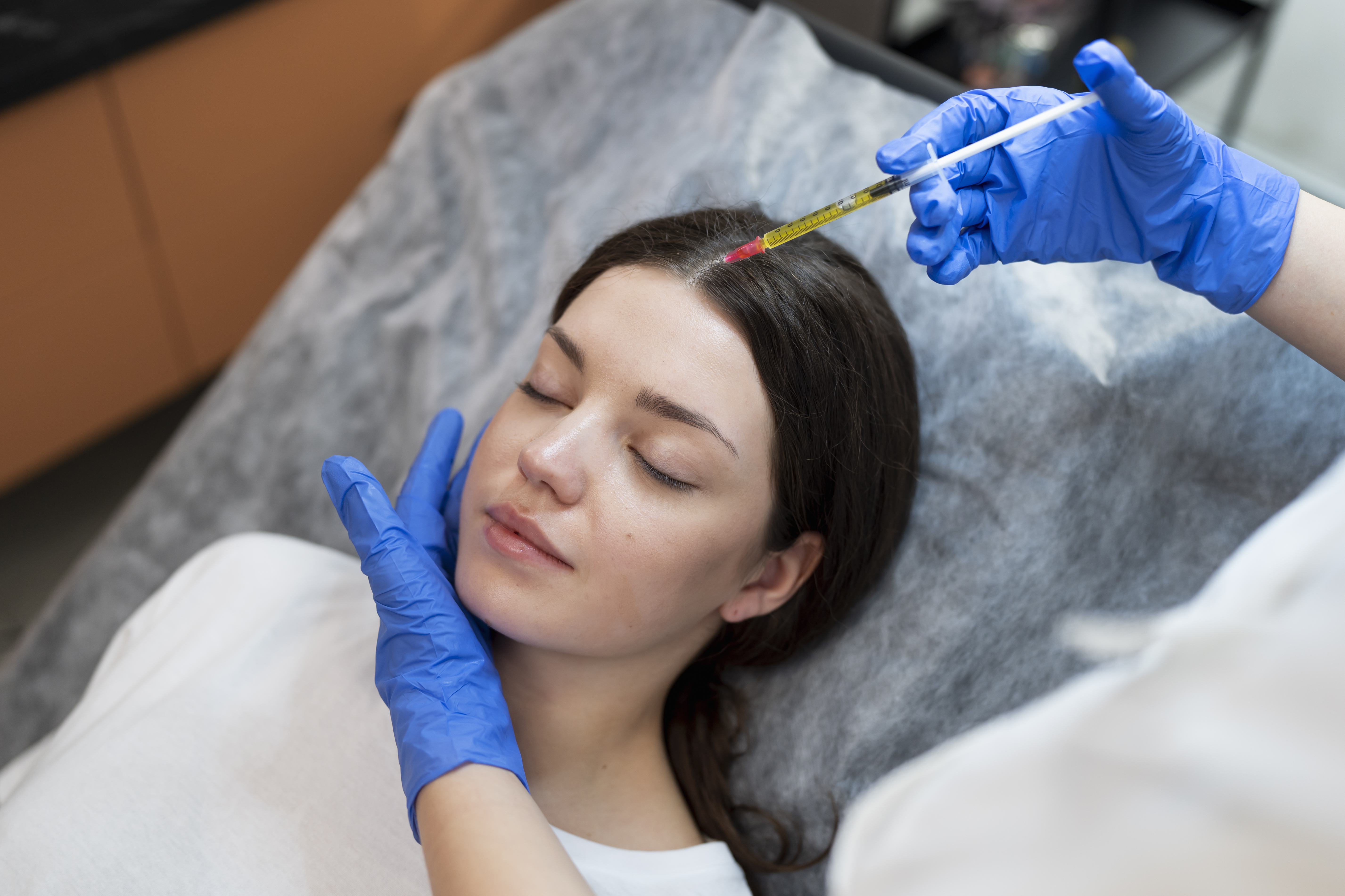
PRP therapy begins by drawing a small amount of blood from the patient at the point of care, which is then processed through centrifugation to isolate the platelets rich in growth factors. These platelets are typically activated and injected into the targeted body area.
This treatment enhances the healing cascade, alleviates pain, leverages naturally inherent therapeutic processes, and promotes tissue regeneration.
PRP is widely used in orthopedics to heal muscles, tendons, joints, and in aesthetics to rejuvenate skin, restore hair growth, support sexual wellness in both men and women, and accelerate wound healing.
- PRP uses the body's blood to promote healing – it is autologous
- It is commonly used in orthopedics, sexual wellness, wound healing, and aesthetics.
- PRP accelerates tissue regeneration, reduces pain, and enhances recovery.
- The properties instigate a body’s natural regenerative cascade; they do not mask but resolve.
- The worst thing that happens is nothing.
Stem cells are derived from various sources, including bone marrow, adipose tissue (also known as fat), umbilical cord blood, and induced pluripotent stem cells (iPSCs). Cord blood-derived mesenchymal stem cells (MSCs) are increasingly preferred in clinical applications due to their high regenerative potential, lower risk of immune rejection, and ethical sourcing.
The History of PRP
PRP therapy originated in the 1970s when researchers discovered the role of platelets in wound healing. Its clinical use began in the 1980s for patients with low platelet counts. The first intraoperative use was during open-heart surgery; soon after, dentists introduced the product to the mainstream with its application in dental implants.
By the 1990s, PRP had become widely used in orthopedics to treat tendon injuries, osteoarthritis, and other musculoskeletal indications.
In the 2000s, PRP expanded into aesthetics through the introduction of the “vampire facial” for skin rejuvenation and hair restoration.
Today, PRP is a trusted treatment across various specialties, including orthopedics, dermatology, urology, podiatry, gynecology, dental surgery, and plastic surgery. Ongoing research continues to explore even more potential applications.
There is rarely a day when PRP isn’t performed on a major athlete and reported on ESPN or in the news. It has become the go-to foundation treatment for many indications.
How PRP is Administered
PRP is a straightforward procedure with four steps:
- Blood Draw: A small amount of blood is drawn from the patient.
- Centrifugation: The blood is processed to isolate the platelets.
- Extraction: The platelet-rich plasma is drawn into a syringe.
- Injection: The PRP is injected into the target area, typically using ultrasound guidance for precision. We recommend activating the PRP with calcium immediately before injection for optimal results.
In the Body:
- All PRP goes through three stages: Inflammatory, Proliferative, and Remodeling.
- Collagen production is stimulated, improving tissue strength.
- Blood flow is enhanced through the formation of new vessels. (angiogenesis and neovascularization)
- The body’s healing cascade involves the release of cytokines, growth factors, and macrophages, which are directed to the targeted area.
PRP in Clinical Practice
PRP is used for various indications, from skin rejuvenation to joint healing. For example:
- Skin Rejuvenation: PRP improves skin texture, tone, and tightness while thickening the epidermis. It is an excellent solution for an underserved market: the back of the hands. Numerous studies have demonstrated exceptional results when treating diabetic foot ulcers.
- Hair Restoration: Stimulates hair growth in areas affected by thinning or balding.
- Orthopedic Healing: Supports recovery from muscle, ligament, and tendon injuries.
- Sexual wellness by improving blood flow in the affected areas for men or women; excellent results are reported when treating women for incontinence with fast improvement.
Efficacy:
- Hair Restoration: An improvement in density and thickness is typically observed within 5 weeks, with a noticeable improvement evident in 3–6 months.
- Aesthetic Treatments: Visible skin improvements occur very quickly when the NanoPen® is used, resulting in durable volume increases in as little as 4 weeks.
- Orthopedic Patients: Modalities such as an ESWT device or cold laser quickly demonstrate improvements. Depending on the severity of the injury, pain relief and improved mobility can be achieved after a single session, with durable results.
- Sexual Wellness: For men experiencing erectile dysfunction, a PRP injection into the corpus cavernosum in concert with a vacuum pump will make an impact in under 14 days. For women, FSD is addressed quickly, and incontinence is often resolved in under 21 days with Kegel exercises.
- Wound Healing: Numerous studies have demonstrated that PRP is an effective treatment option for diabetic foot ulcers.
PRP vs. Other Treatments
- Market PRP heavily with no regulatory issues, compared to cell therapy, which is scrutinized.
- The source is the patient's blood versus bone marrow, fat, or a Synthetic substance.
- The mechanism boosts natural healing, repairs tissue, and adds volume and hydration.
- Use Cases: Hair, Skin, Joints, plantar fasciitis, Sexual Wellness, DFU, Burns, Scar Revision, Advanced Degeneration, Wrinkles, Facial Volume.
- The cost is minimal compared to other modalities.
- Minimal Downtime, if any.
- The worst outcome is that nothing changes.
Key Considerations:
- Use only an FDA-cleared PRP system that has been proven safe and effective.
- Your PRP kit should yield at least 3.5 times the platelet concentration.
- Ensure proper clinician training to ensure optimal results.
Additional Resources:
Orthopedics:
- Lateral Epicondylitis (Tennis Elbow): High-quality evidence supports leukocyte-rich platelet-rich plasma (LR-PRP) injections for treating lateral epicondylitis, resulting in significant pain reduction and functional improvement. PubMedCentral
- Knee Osteoarthritis: Leukocyte-poor platelet-rich plasma (LP-PRP) injections have shown promise in alleviating pain and improving function in patients with knee osteoarthritis, PMC , MDPI
- Patellar Tendinopathy and Plantar Fasciitis: Moderate evidence supports PRP injections for these conditions, offering potential pain management and tissue healing benefits. PMC
- Rotator Cuff Tendinopathy and Hip Osteoarthritis: There is sufficient evidence to recommend the routine use of PRP for these conditions. PMC Article on Rotator Cuff Tendinopathy , ScienceDirect on Rotator Cuff Tears, PMC Article on Hip Osteoarthritis , BMC Musculoskeletal Disorders on Hip OA
Aesthetics and Dermatology:
- Hair Restoration: PRP has been beneficial for treating alopecia areata and androgenetic alopecia and improving hair density and thickness. A regimen of three treatments six weeks apart is typically recommended, followed by annual maintenance sessions. Cleveland Clinic
- Skin Rejuvenation: PRP injections can enhance skin texture, tone, and elasticity, contributing to facial aesthetic improvements.
- Patient Selection: PRP therapy is generally considered safe, as it utilizes autologous components. However, patients with platelet dysfunction syndromes or active infections should be evaluated cautiously.
- Pre-Procedure Recommendations: Discontinuation of anti-inflammatory medications and blood thinners may be advised before the procedure to optimize outcomes. Northeast Knee & Joint Institute.
- Injection Protocol: PRP is typically administered as a single injection; however, some protocols suggest a series of injections over several weeks, depending on the condition and its severity. Kaiser Permanente Washington
Platelet-Rich Plasma (PRP) Therapy
UnitedHealthcare Medicare Advantage Medical Policy
This policy outlines coverage guidelines for PRP therapies, including CPT code 0232T for PRP injections. It specifies that PRP injections are non-covered for specific indications, while coverage may be considered for chronic non-healing wounds under certain conditions and in accordance with reimbursement guidance.
Post-Procedure Care
Activity Modification
To facilitate healing, patients may be advised to rest the treated area briefly, followed by a gradual return to activity. For orthopedic purposes, a good rule of thumb is maintaining a 40% activity level for 4 weeks, increasing to 50% at 5 weeks, and returning to full activity at 6 weeks.
Pain Management
Mild soreness at the injection site is common. Over-the-counter pain relievers may be recommended, with the avoidance of NSAIDs, as they can diminish cell signaling. This approach is counterintuitive to our objective, as we want the body to heal itself.
Safety Considerations
Adverse Effects: PRP injections are generally low-risk. Potential side effects include soreness, bruising, or swelling at the injection site. Serious complications are rare due to the autologous nature of the therapy.
Source: Johns Hopkins Medicine
Regulatory and Standardization Aspects
Clinical Guidelines: While PRP therapy shows promise, variations in preparation techniques and injection protocols exist. Standardized guidelines and further research are necessary to establish optimal practices and indications for using PRP.
Understanding the Science Behind PRP
PRP’s therapeutic effects are driven by three core biological components: platelets, growth factors, and cytokines. These elements work together to repair tissue, stimulate cell growth, and control inflammation.
- Platelets: These tiny cells in the blood play a crucial role in clotting and initiating the healing process. They release growth factors that repair tissues and stimulate the production of collagen.
- Growth Factors: Proteins such as PDGF, TGF-β, and VEGF promote tissue regeneration, stimulate cell growth, and enhance collagen production.
- Cytokines: These proteins regulate the immune system and inflammation, ensuring a fast and efficient healing process.
When PRP is injected into a targeted area, the platelets release growth factors and cytokines, which promote tissue regeneration. This leads to increased collagen production and improved blood flow, allowing the body to heal more effectively and reducing inflammation. The healing cascade is also stimulated, prompting the bone marrow to release mesenchymal stem cells (MSCs) targeting the damaged area. Macrophages then carry away the scar tissue and debris through the lymphatic system.
How It Works
- Collagen Production: PRP stimulates collagen growth, facilitating the repair and regeneration of skin, tendons, and other tissues.
- Improved Blood Flow: New blood vessels form, enhancing circulation to the treated area, thereby accelerating the healing process.
- Reduced Inflammation: Cytokines help regulate inflammation, making the recovery process smoother and more efficient.
The Role of PRP in Medicine
PRP is a game-changer in both regenerative and aesthetic medicine. It provides a noninvasive, effective alternative to surgery for soft tissue injuries, pain relief, and tissue regeneration.
In addition to orthopedic uses, PRP has gained widespread popularity in aesthetics, offering treatments for skin rejuvenation, sexual wellness, hair restoration, and general anti-aging.
Applications of PRP
- Regenerative Medicine: Heals muscle, tendon, and joint injuries and can treat osteoarthritis and tendonitis.
- Aesthetic Medicine: Improves skin health, restores hair, and reduces fine lines and wrinkles.
- Sexual Wellness: Addresses ED, FSD, and incontinence.
- Wound Healing: Great results for DFUs and scar revision.
Choosing the Right PRP System
Not all PRP systems are the same. The quality of platelet concentration varies by equipment; therefore, clinicians must select FDA-approved systems that have proven to improve platelet concentration. Additionally, proper training is essential for achieving optimal results.
Key Considerations for Clinicians:
- FDA Approval: Ensure the system is FDA-cleared for safety and efficacy.
- Training: Clinicians should receive thorough training on correctly using the equipment.
- Platelet Concentration: PRP preparation should have a higher concentration of platelets than normal whole blood. A typical target for therapeutic PRP is 3 to 5 times the baseline platelet concentration in blood. Higher concentrations can yield better therapeutic results, thereby enhancing the healing process.
Practical Information
The platelet concentration is one of the most critical variables when using automated centrifugation systems to separate platelets from whole blood. Look for equipment that allows for precise control of platelet yield to ensure consistency and uniformity.
Recent Research
Studies suggest higher platelet concentrations are associated with better clinical outcomes in orthopedic and dermatological applications following treatment. PubMed+
Red Blood Cells (RBCs)
RBCs in PRP should be minimized, as excessive RBCs may contribute to increased pain, inflammation, and complications at the injection site. A study demonstrated that LP (leukocyte-poor) PRP was significantly more effective when administered intra-articular injection.
Practical Information
The goal is to isolate PRP with minimal RBCs, resulting in a cleaner and more effective preparation. The lower the RBC contamination, the better.
Research
High RBC levels can interfere with releasing growth factors and the overall healing response. (Follman et al., 2019, PubMed)
pH
The pH of PRP should ideally range between 6.8 and 7.4. Deviation from this range can affect platelets’ viability and ability to release growth factors effectively.
Practical Information
pH levels should be tested after preparation to ensure they are within the ideal range. Some PRP systems include pH monitoring to adjust as needed. However, FDA test results should be reviewed before purchase.
Latest Research
pH changes can cause platelet degranulation, thereby affecting the regenerative potential of the treatment. (Jin et al., 2018, PubMed)
Hematocrit (HCT)
HCT represents the percentage of blood volume occupied by red blood cells (RBCs). An ideal hematocrit level in PRP is generally low. High HCT values can negatively affect the PRP’s regenerative properties.
Practical Information
If the HCT is too high, the PRP preparation may have a thicker consistency, impairing its ease of injection and causing patient discomfort.
Latest Research
HCT should be monitored, as higher HCT can result in a lower platelet yield, reducing the overall effectiveness of the treatment.
Link to Study: The Effect of Platelet-Rich Plasma Formulations and Blood Products on Synoviocyte Activation – PMC
White Blood Cells (WBC)
WBCs can sometimes be beneficial, as they may contribute to the inflammatory phase of wound healing. However, excessive WBCs may lead to prolonged inflammation and other complications.
Practical Information
WBC levels should be minimized, as excessive leukocytes can lead to unwanted side effects, such as infection or prolonged inflammation.
Latest Research
Some studies suggest that WBCs in PRP can be counterproductive, leading to chronic inflammation. (Cakir et al., 2017, PubMed)
Competitive Products
When evaluating competitive PRP systems, it’s essential to assess how efficiently the system isolates platelets, RBCs, and WBCs. Look for systems that precisely control the centrifugation process, allow customizable platelet concentrations, and ensure minimal RBC contamination. The latest PRP devices use advanced filtration methods to achieve these results.
Real-World Applications and Results: Patient Case Studies
Case Study 1: Orthopedic – Chronic Musculoskeletal Injury
Patient Profile:
- Age: 42
- Gender: Male
- Occupation: Professional tennis player
- Medical History: Chronic tennis elbow for over 2 years, unresponsive to physical therapy and corticosteroid injections.
Treatment Process:
After a consultation and ultrasound imaging, the patient was diagnosed with lateral epicondylitis (tennis elbow) and recommended PRP therapy.
The patient prepared by discontinuing all NSAIDs for 14 days.
Blood was drawn, processed in a commercially available kit to concentrate the platelets, and injected into the injured tendons of the elbow under ultrasound guidance and local anesthesia via a peri-articular methodology.
The patient was advised to rest for two weeks post-treatment, avoid ice, and continue regular physical activity.
Outcomes:
- Initial Assessment (Day 14): Patient reported a noticeable reduction in pain.
- Follow-up (Day 32): Patient resumed light training with improved grip strength, range of motion, and minimal discomfort.
- Follow-up (Day 60): Patient was pain-free and fully recovered, with no further issues during play, and returned to professional matches.
Conclusion:
PRP therapy effectively reduced inflammation and promoted tendon healing, enabling the patient to return to professional play quickly.
An annual booster program has been recommended.
Case Study 2: Knee – Osteoarthritis
Patient Profile:
- Age: 58
- Gender: Female
- Occupation: Office worker
- Medical History: Diagnosed with severe knee osteoarthritis for over 5 years, with significant pain and limited mobility despite two corticosteroid injections, oral NSAIDs, and physical therapy.
Treatment Process:
After a consultation and MRI imaging, the patient was diagnosed with advanced knee osteoarthritis. PRP therapy was recommended. Blood was drawn and processed to concentrate the platelets. Calcium chloride (5% of the injectable yield) was added to activate the platelets and injected intraarticularly into the knee joint, utilizing ultrasound guidance. The patient was advised to avoid heavy physical activity for two weeks following the procedure.
Outcomes:
- Initial Recovery: Two weeks post-treatment, the patient reported a noticeable decrease in pain and swelling. The range of motion had increased by 25%, and exercise levels had also increased accordingly.
- Follow-up (30 Days): Thirty days after treatment, the patient experienced improved knee function, significantly increased mobility, and reduced pain during daily activities. Increased levels of exercise included walking up to 5 miles per day and light jogging.
- Follow-up (90 Days): 90 days after the initial assessment, the patient could jog regularly, began practicing yoga 5 days a week, and participated in a light exercise class. The pain was almost non-existent, and all NSAID use was stopped, allowing her to engage in more physical activities.
Conclusion:
PRP therapy for knee osteoarthritis effectively alleviated pain, improved joint function, and significantly enhanced the patient’s quality of life, enabling her to return to her normal daily activities without the need for surgery.
Case Study 3: Shoulder – Rotator Cuff Injury
Patient Profile:
- Age: 45
- Gender: Male
- Occupation: Construction worker
- Medical History: Rotator cuff tendinopathy for 6 months, causing persistent shoulder pain and weakness, particularly during overhead motions. The patient had been unresponsive to corticosteroid injections and physical therapy.
Treatment Process:
After a clinical examination and MRI, the patient was diagnosed with chronic rotator cuff tendinopathy. PRP therapy was recommended in concert with ESWT. Blood was drawn and processed to concentrate the platelets. Calcium gluconate was administered to activate the platelets. They were then injected periarticular into the shoulder joint area, and the affected tendons were bathed under ultrasound guidance and local anesthesia. Six ESWT treatments were performed over the following 21 days, with 3-day intervals between sessions. The patient was advised to rest for two weeks post-treatment, with a gradual return to rehabilitation exercises.
Outcomes:
- Initial Recovery: Two weeks post-treatment, the patient reported a noticeable decrease in pain and swelling. The range of motion had increased by 25%, and exercise levels had also increased accordingly.
- Follow-up (21 Days): At 21 days, the patient resumed part-time work and physical therapy, exhibiting improved shoulder strength and an increased range of motion.
- Follow-up (65 Days): At 65 days, the patient returned to his normal full-time work activities, including lifting and overhead motions, with no lingering discomfort.
Case Study 4: Aesthetic – Hair Restoration for Androgenic Alopecia
Patient Profile:
- Age: 30
- Gender: Female
- Medical History: Androgenic alopecia for 5 years, noticeable hair thinning at the crown and temples.
Treatment Process:
A comprehensive hormone panel blood test was performed, and no significant deficiencies were identified. PRP therapy was recommended to address hair thinning. A ring block was performed, blood was drawn and processed to isolate platelets, and calcium was added and injected into the scalp, targeting the areas with thinning hair. The injections were performed under local anesthesia, and a microneedle device infused PRP topically post injections; the patient was instructed to avoid washing her hair for 8 hours post-treatment.
Outcomes:
- Initial Recovery: Mild tenderness in the treated areas, with no significant discomfort.
- Follow-up (21 Days): The patient noticed small, fine hair follicles regrowing in the treated areas and reduced hair loss during showers.
- Follow-up (42 Days): The patient experienced thicker, healthier hair, particularly around the crown and temples. She was highly satisfied and opted for a second round of PRP therapy. An identical treatment process, including the microneedle procedure, was performed.
- Follow-up (90 Days): The patient had noticeable hair growth in her temples and widow’s peak and significant improvement in coverage in her crown. She experienced thicker, healthier hair, particularly on her head and forehead. She committed to 2 annual booster treatments per year.
Conclusion:
PRP therapy significantly enhanced hair density and texture, promoting hair regrowth in a patient with androgenic alopecia.
Case Study 5: Aesthetic – Skin Rejuvenation (Vampire Facial)
Patient Profile:
- Age: 50
- Gender: Female
- Medical History: Mild fine lines, age spots, and skin dullness; seeking a non-invasive treatment for skin rejuvenation.
Treatment Process:
The patient underwent a “vampire facial,” in which PRP was used for skin rejuvenation. After drawing and processing the blood, calcium was added to activate the plasma. She then received twelve intradermal platelet-rich plasma injections, allowing the PRP to reach deeper skin layers and stimulate collagen production. This was followed by 1 mL of PRP infusion into the epidermis via a microneedle device. The patient was advised to avoid makeup and the sun for 24 hours and apply moisturizer afterward.
Outcomes:
- Initial Recovery: Mild edema, erythema, and swelling lasted for 2 days, which were alleviated with the use of cooling gels.
- Follow-up (16 Days): The patient reported a significantly improved skin texture, smoother and reduced fine lines and wrinkles.
- Follow-up (45 Days): The patient’s skin demonstrated significant improvement, becoming firmer and more youthful and exhibiting a reduction in age spots and fine lines. Volume boosts in the nasolabial folds were evident.
Conclusion:
The PRP-based vampire facial rejuvenated the skin, enhancing texture and firmness while reducing fine lines with a microneedle device.
Latest Research on PRP
Phases of Wound Healing Enhanced by PRP:
Inflammatory Phase
Role in Healing: This initial phase involves the body’s response to injury, characterized by hemostasis and the clearance of pathogens and debris. It typically lasts between one and three days.
PRP’s Contribution: PRP contains a high concentration of platelets, which release growth factors such as PDGF, TGF-β, and VEGF. These factors recruit and activate neutrophils and macrophages, which are essential for decontaminating the wound site and secreting cytokines that orchestrate subsequent healing stages.
Proliferative Phase
Role in Healing: This phase focuses on tissue formation, including angiogenesis, collagen deposition, and epithelialization, typically lasting between 21 and 28 days.
PRP’s Contribution: The growth factors in PRP stimulate fibroblasts to synthesize collagen and extracellular matrix components, facilitating tissue repair. Additionally, PRP enhances keratinocyte migration, promoting re-epithelialization. Studies have shown that PRP application accelerates wound closure and improves tissue regeneration.
Remodeling Phase
Role in Healing: The final phase, which can last up to a year, involves maturing and strengthening the newly formed tissue.
PRP’s Contribution: By providing a sustained release of growth factors, PRP supports the remodeling of collagen fibers, thereby improving tensile strength and tissue integrity. Research indicates that PRP can modulate the wound-healing process by balancing the microenvironment, promoting appropriate wound healing.
Recent Research and Findings
Study Title:
Platelet-Rich Plasma and its Role in Wound Healing and Soft Tissue Regeneration: A Review
Published in:
Study Focus:
This study examines the impact of PRP on collagen remodeling and its role in the remodeling phase of wound healing.
PRP’s Impact on Tendon Healing
Study:
“Effect of a Single Versus Serial Platelet-Rich Plasma Injection on the Healing of Acute Achilles Tendon Injuries in Rats” (2024)
Published in:
BioMed Central
Findings:
This study examined the timing and frequency of platelet-rich plasma (PRP) injections in tendon healing. It concluded that early administration of PRP enhances tendon healing, while multiple injections after the initial healing phase might impede recovery. A single PRP injection during tendon repair surgery was more effective than multiple injections.
PRP Concentration Effects on Wound Remodeling
Study:
“Concentration-Dependent Effect of Platelet-Rich Plasma on Keratinocyte and Fibroblast Wound Healing Potential” (2014)
Published in:
ScienceDirect – Platelet-rich plasma in orthopedic sports medicine
Findings:
This study investigated the impact of varying PRP concentrations on wound healing. A 10% PRP concentration promoted wound remodeling, whereas a 20% concentration enhanced inflammation and collagen deposition. These results underscore the importance of optimizing PRP concentration for therapeutic applications.
Link:
ScienceDirect – Platelet-rich plasma in orthopedic sports medicine
The Science Behind PRP
What’s in PRP?
- Platelets: Blood cell fragments essential for clotting and healing.
- Growth Factors: Proteins like PDGF, TGF-β, VEGF, and EGF that drive tissue repair.
- Cytokines: Small proteins that help regulate inflammation and immune response.
How PRP Works
When injected into an area of injury or aging, PRP releases growth factors that:
- Stimulate collagen production.
- Encourage angiogenesis and neovascularization — the formation of new blood vessels.
- Reduce inflammation and accelerate healing.
In simple terms: PRP harnesses the body’s inherent healing cascade to naturally promote self-healing, rejuvenate tissues, and accelerate recovery rather than merely mask pain or discomfort.