Diagnostic Ultrasound
- Home
- Diagnostic Ultrasound
Introduction to diagnostic ultrasound
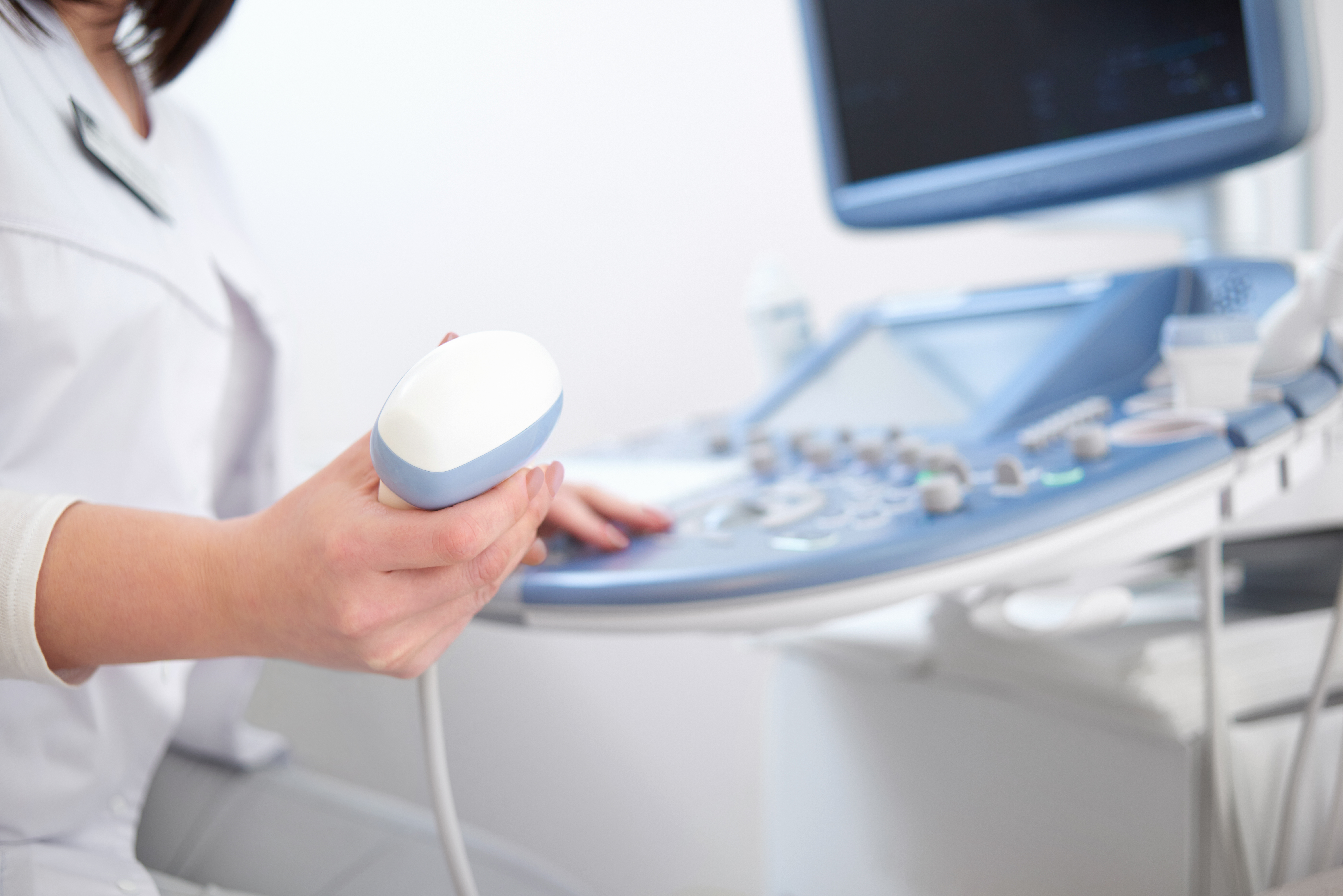
Diagnostic ultrasound, also known as sonography, is a noninvasive imaging technique that utilizes high-frequency sound waves to produce real-time images of internal body structures. Unlike X-rays or CT scans, ultrasound does not use ionizing radiation, making it a safer option, especially for sensitive groups like pregnant women and children.
Ultrasound Applications
Ultrasound is widely used across various medical specialties, including:
- Obstetrics: Monitoring fetal development, detecting abnormalities, and evaluating placental health.
- Cardiology: Assessing heart function, measuring blood flow, and visualizing the heart’s chambers and valves.
- Musculoskeletal: Imaging muscles, tendons, and joints to diagnose injuries like sprains, tears, and inflammation.
- Abdominal Imaging: Ultrasound is commonly used to visualize the liver, kidneys, and gallbladder. It’s a non-invasive tool for diagnosing liver disease, kidney stones, and gallstones. (source)
- Vascular: Used for evaluating blood flow, detecting blockages, and diagnosing conditions like DVT and PAD. Doppler ultrasound helps assess blood circulation in veins and arteries. (source)
- Thyroid: Ultrasound assists in evaluating thyroid nodules and distinguishing between benign and malignant masses. It’s key in guiding biopsies for further examination. (source)
- Breast: Ultrasound helps differentiate between benign and malignant breast lesions, providing valuable support in interpreting mammography results. It’s also used to guide biopsies. (source)
- Procedure Guidance: Ultrasound provides real-time imaging for guiding procedures such as biopsies, injections, and fluid drainage, ensuring precision and reducing risks. (source)
What to Look for in a Competitive Product
- Image Resolution: Higher-resolution systems will provide more detailed and precise images.
- Portability: Essential if you need ultrasound in different settings, such as emergency rooms or remote areas.
- Ease of Use: Look for intuitive, user-friendly systems with simple interfaces and adjustable settings.
- Advanced Features: Doppler imaging for analyzing blood flow or 3D imaging for obstetrics can add significant value.
Practical Tip
Ensure the system can accommodate multiple diagnostic needs, making it a versatile tool across various specialties.
Science of Diagnostic Ultrasound
Ultrasound emits high-frequency sound waves, typically 2 to 18 MHz, through a transducer. These sound waves interact with tissues, and the resulting echoes are captured by the transducer and transformed into visual images.
Different tissues reflect sound waves to varying degrees, which allows the ultrasound machine to distinguish between muscle, fat, and organs.
Real-Time Imaging and Interpretation of Ultrasound Data
Ultrasound’s most significant advantage is its ability to provide real-time images. This feature is particularly valuable for guiding procedures like needle biopsies, injections, and surgeries. With real-time imaging, clinicians can make immediate decisions during procedures, improving precision and outcomes.
What to Look for in a Competitive Product:
- Choose systems that deliver high-quality resolution for still images and video
- Look for color Doppler imaging and tissue elasticity mapping for precise information
- Particularly important for specialties like cardiology and oncology
Real-World Example
In cardiology, high-quality Doppler ultrasound systems can precisely assess blood flow patterns, helping clinicians diagnose heart diseases, valve issues, and congenital disabilities in infants.
Historical Development of Diagnostic Ultrasound
Evolution from Early Diagnostic Tools to Modern High-Resolution Imaging Systems
Ultrasound technology began in the 1950s with large, basic machines that detect tissue abnormalities. Over time, advancements introduced higher-frequency sound waves, Doppler imaging, and 3D/4D capabilities, significantly improving resolution and diagnostic accuracy.
Key Advancements in Portability and Precision
Developing portable ultrasound systems was a game-changer, particularly in emergency and point-of-care settings.
Handheld ultrasound units have enabled imaging in nontraditional settings, such as ambulances and remote areas. They provide quick access to diagnostic imaging without requiring patients to be transported to a hospital for treatment.
What to Look for in a Competitive Product
When selecting ultrasound equipment, consider portability and image precision. Modern ultrasound systems are available in various sizes, ranging from full-sized hospital units to portable, handheld devices. Select the one that balances high-quality imaging and ease of transport, primarily if you work in mobile or urgent care settings.
- Batter Life: Ensure the ultrasound device has long-lasting battery life, especially for portable or handheld units in mobile or urgent care settings. A longer battery life enables continuous usage without frequent recharging, enhancing efficiency.
- Artificial Intelligence (AI): Look for ultrasound equipment that integrates AI for enhanced imaging capabilities. AI can assist in automatic image analysis, improve diagnostic accuracy, and streamline workflow by reducing the need for manual interpretation.
DICOM (Digital Imaging and Communications in Medicine)
Ensure the ultrasound system supports DICOM compatibility for easy integration into hospital or clinic systems. DICOM enables the seamless storage, retrieval, and sharing of medical images, ensuring efficient patient data management.
Latest Research
A recent study in the Journal of Ultrasound in Medicine highlighted the importance of portable ultrasound machines in enhancing access to healthcare in low-resource regions while maintaining diagnostic accuracy.
Mechanisms of Action
How Ultrasound Waves Penetrate Tissues and Create Visual Images
When ultrasound waves are transmitted into the body, they travel through tissues and reflect, depending on the tissue type. The ultrasound system processes these returning echoes to generate detailed images visually representing internal structures. Differences in tissue density, such as between solid organs, muscles, and fluids, help form clear images.
Importance of Doppler Ultrasound for Assessing Blood Flow
A Doppler ultrasound is a specialized technique to measure blood flow within vessels. By tracking the frequency shift of sound waves bouncing off moving red blood cells, Doppler ultrasound provides critical information about the speed and direction of blood flow, essential for diagnosing conditions such as arterial blockages and venous insufficiencies.
What to Look for in a Competitive Product: Choose a system with advanced Doppler features. High-end ultrasound systems offer color Doppler imaging, which visually color-codes blood flow, making it easier to interpret results quickly.
Real-World Example: Doppler Ultrasound in Obstetrics
Doppler ultrasound is commonly used in obstetrics to monitor blood flow in the umbilical cord, assess fetal health, and detect complications related to placental circulation. Here are patient case studies that demonstrate how Doppler ultrasound is applied in clinical settings to ensure the health of both mother and baby.
Obstetric Case Study: Monitoring Blood Flow in the Umbilical Cord
Patient Profile:
- Name: Rachel
- Age: 32
- Gender: Female
- Medical History: First-time pregnancy, no significant medical history.
- Gestational Age: 28 weeks
Treatment Process
Rachel underwent a routine ultrasound to monitor fetal health. The obstetrician recommended a Doppler ultrasound due to her family history of hypertension. During the procedure, high-frequency sound waves were used to measure the blood flow through the umbilical cord. The results were analyzed to assess the speed and direction of blood flow, ensuring that the fetus received adequate oxygen and nutrients.
Outcomes
Week 1: The Doppler ultrasound revealed normal blood flow in the umbilical cord, indicating that the fetus was receiving sufficient blood supply.
Follow-Up: At the 32-week follow-up, a subsequent Doppler ultrasound revealed no signs of placental insufficiency or abnormal blood flow, confirming that both mother and baby were doing well.
Findings
The Doppler ultrasound effectively monitored Rachel’s pregnancy, ensuring the proper functioning of the umbilical cord and placental circulation. The procedure provided peace of mind and confirmed no immediate concerns for fetal health.
Obstetric Case Study: Assessing Fetal Health and Placental Circulation
Patient Profile:
- Name: Laura
- Age: 36
- Gender: Female
- Medical History: Previous miscarriage, high-risk pregnancy due to age, and underlying conditions, including gestational diabetes.
- Gestational Age: 24 weeks
Treatment Process
Laura’s pregnancy was classified as high-risk due to her medical history and age. To assess the health of her fetus and the function of the placenta, a Doppler ultrasound was performed. The procedure measured the blood flow through the umbilical cord and the uterine arteries, evaluating the blood supply to the placenta and fetal organs.
Outcomes
Week 1: The Doppler ultrasound showed normal blood flow to the fetus and placenta, with no signs of restricted blood flow or fetal distress.
Follow-Up: At 30 weeks, another Doppler ultrasound revealed that the placental blood flow remained optimal, a positive indicator of continued fetal health.
Final Outcome: By 38 weeks, Laura successfully delivered a healthy baby, and the Doppler ultrasound provided vital information throughout the pregnancy, ensuring no complications arose from placental or fetal circulation issues.
Findings
The Doppler ultrasound played a crucial role in monitoring Laura’s high-risk pregnancy. It helped track placental health and fetal well-being, enabling early intervention if necessary and ensuring a successful pregnancy outcome.
Obstetric Case Study: Detecting Placental Complications
Patient Profile:
- Name: Megan
- Age: 40
- Gender: Female
- Medical History: History of preeclampsia in a previous pregnancy, currently pregnant with twins.
- Gestational Age: 26 weeks
Treatment Process
Given Megan’s history of preeclampsia, her obstetrician ordered a Doppler ultrasound to assess placental circulation and monitor fetal health. The procedure focused on measuring blood flow through the umbilical cords of both babies and the uterine arteries to detect early signs of complications, such as intrauterine growth restriction (IUGR) or preeclampsia-related placental insufficiency.
Outcomes
Week 1: The Doppler ultrasound results showed reduced blood flow in the uterine artery, indicating a slight risk of developing preeclampsia. However, the blood flow to the fetuses was within normal ranges.
Follow-Up: At 30 weeks, a second Doppler ultrasound was performed. The results showed that the blood flow to Baby A was slightly restricted, prompting further monitoring and adjustments to Megan’s care plan.
Final Outcome: By 36 weeks, the second baby was born without complications, but Baby A required a brief stay in the neonatal intensive care unit (NICU) for monitoring. The Doppler ultrasound enabled the early identification of potential complications, facilitating timely interventions.
Findings
The Doppler ultrasound was instrumental in monitoring Megan’s high-risk twin pregnancy. It detected early signs of placental complications, allowing for proactive management and reducing risks to both babies. The procedure ensured optimal outcomes despite the challenges posed by her medical history.
Case Study 4: Ultrasound-Guided Acromioclavicular (AC) Joint Injection for Shoulder Pain Relief
Patient Profile:
- Age: 65
- Occupation: Manual laborer
- Gender: Female
- Medical History: Persistent right shoulder pain unresponsive to conservative treatments
Clinical Presentation
The patient presented with chronic right shoulder pain, mainly localized over the AC joint. Despite undergoing physical therapy and taking nonsteroidal anti-inflammatory drugs (NSAIDs), she experienced minimal relief. The pain significantly impacted her ability to perform daily tasks, particularly those that required overhead movements.
Diagnostic Approach
Physical examination revealed tenderness over the AC joint and limited shoulder range of motion. Imaging studies, including X-rays, confirmed signs of AC joint arthritis. An ultrasound examination was performed to further assess the condition and guide treatment. The examination identified inflammation and degeneration within the AC joint.
Treatment Intervention
An ultrasound-guided injection of the AC joint was administered, consisting of a local anesthetic and corticosteroid to reduce inflammation and alleviate pain. Using ultrasound guidance ensured precise needle placement, enhancing the effectiveness of the injection and minimizing potential complications.
Outcomes
One month after the injection, the patient reported pain relief and had regained full shoulder mobility. She returned to her manual labor job without discomfort and experienced no adverse effects from the procedure.
Conclusion
This case demonstrates the efficacy of ultrasound-guided AC joint injections in providing significant pain relief and functional improvement for patients with AC joint arthritis who are unresponsive to conservative treatments. The precision offered by ultrasound guidance significantly contributes to the success and safety of the procedure.